Technology
- 3-Dimensional CAT Scan Machine
- Digital X-Rays
- CEREC CAD/CAM
- Intraoral Scanner and Camera
- Computer Guided Surgery
- Microscopic Surgery
- Laser and Piezoelectric Surgery
- EKG Monitoring
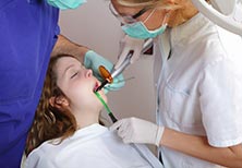
Dentistry is a technologically advanced, rapidly evolving
field. New discoveries and developments are constantly raising the bar on what
is the very best available. Because we pride ourselves in offering nothing but
the best, we regularly update our facility with newer, better equipment. As Dr.
Jain, founder of Center for Implant Dentistry, said, "I would not buy myself a
Mercedes car, but I buy the Mercedes of dental equipment for my office."
3-Dimensional CAT Scan Machine


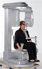 This is by far the most important piece of equipment in our
office. Although some practices attempt to place implants using only 2D
(standard x-ray) images, we believe that is a disservice to patients. Accurate
diagnostics and precision planning are keys to healthy, successful treatment.
This is by far the most important piece of equipment in our
office. Although some practices attempt to place implants using only 2D
(standard x-ray) images, we believe that is a disservice to patients. Accurate
diagnostics and precision planning are keys to healthy, successful treatment.
This machine generates high-definition, 3D digital images,
providing your doctor with precision anatomical information about the maxillofacial
and oral structures, while lessening your exposure to radiation. Simply put, it
gives us the ability to see what we need to see BEFORE we begin surgery, thus
avoiding complications and surprises.
Digital X-Rays
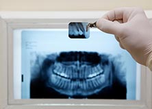
Old-fashioned film-based x-rays are quickly being replaced
by digital machines in many dental offices. As leaders in advanced technology,
we have been using this technique for some time, and found it to be quite
beneficial to the patient.
The process is much more comfortable than traditional
x-rays, and the digital image is instantly transferred to specialized software.
Once the image has been attained; we can use the software to look very closely
at specific areas of the teeth and surrounding structures. It is also much
healthier than traditional x-rays, exposing the patient to 80 percent less
radiation.
CEREC CAD/CAM
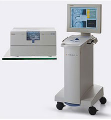
CEREC technology allows us to provide dental restorations in a day,
with no goopy impressions or a temporary. We can create a variety of
restorations right here in our office, during your appointment. CEREC may be
used for:
Inlays, Onlays, Full Crowns, Porcelain Veneers
Inlays, Onlays, Full Crowns, Porcelain Veneers
CEREC restorations are made of beautiful, durable porcelain,
very similar what is used in a dental laboratory. The important difference in
materials is that CEREC porcelain is compressed, giving it superior strength to
the layered porcelain that comes from a lab. There is no goopy, uncomfortable
tray to bite down on, and no need to wear a temporary restoration. Despite the
many other advantages of CEREC, most patients consider the greatest benefit to
be the convenience of single-visit dentistry.
The process of restoring a tooth
with CEREC is very simple and straightforward, but it is technique sensitive. Our
team is highly trained and skilled in the use of all technology in our office,
including CEREC. The procedure is completed in just a few steps:
All decayed, diseased tooth material is removed. The
tooth is shaped as needed for optimal retention of the restoration.A fine
powder is applied, which allows the digital imaging machine to capture detailed
data. A digital impression is taken, which is similar to photographing the tooth –
no goop, and no discomfort.The image
is displayed on a computer screen, and your doctor uses specialized
software to design your new restoration while you watch. The
data is sent to the CEREC milling machine, which sculpts your restoration
from a solid block of porcelain, which takes approximately 15 minutes. The
fit and color of the restoration are verified, and it is cemented in
place. That’s it! The process is
completed in about an hour.
Intraoral Scanner and Camera
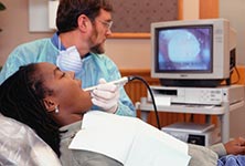
This is a valuable diagnostic tool, which allows both you
and your doctor to see exactly what is happening in your mouth. The camera is
very small, allowing us to view teeth clearly from various angles, without
causing patient discomfort. Images are displayed in amazing detail on a large
screen, and we can zoom in on areas of particular concern.
In addition to precision diagnostics, the intraoral camera
allows us to show you exactly what we are seeing, and why we are recommending a
specific treatment. Because these images provide proof of damage or other
dental problems, they can be quite helpful in achieving treatment approval from
your insurance company.
Computer Guided Surgery

Computer tomography (CT or CAT) was initially introduced to dentistry in the late 1980s, raising the bar on dental implant treatment. This revolutionary technology provided an unprecedented opportunity for the accurate evaluation of anatomic structures with detailed accuracy. CT devices and applications evolved rapidly, as clinicians discovered innovative ways to put this technology to work for the patient.
Incorporating the highly detailed imagery of CT scanning, along with specialized software, the surgery can be performed virtually, before the dentist ever touches the patient. This effectively takes the guesswork out of the actual procedure. It also allows for the fabrication of the final restoration prior to implant placement.
Microscopic Surgery

We use a Surgical Operating Microscope (SOM) to ensure precision accuracy during oral surgeries. It allows your dentist to clearly see fine details of the soft tissue, tooth, and bone structures. Often, the naked eye alone cannot see superficial fractures, subtle changes, or the earliest signs of disease and decay.
With the use of microscopic technology, we can deliver more precise diagnostics, accurate surgical planning, and efficient procedures. This greatly reduces the potential for error or imperfection, and improves overall efficiency of dental care.
Laser and Piezoelectric Surgery
Traditional surgical techniques utilize a scalpel, which is a small, sharp knife. Incisions, like any other cuts, tend to bleed, and they take time to heal. Fortunately, thanks to today’s advanced technology, a scalpel is rarely necessary for oral surgery. We can often complete even complex implant placement procedures with no cutting, and little to no post procedure soreness.
Laser surgery utilizes the power of concentrated, filtered light energy to remove soft or hard tissue with precision. Because it is not “cutting,” it does not irritate surrounding tissues. It generates thermal energy, which has a cauterizing effect, eliminating the need for stitches, and virtually eliminating bleeding.
Piezoelectric surgery is a revolutionary new option for sculpting hard tissue. Rather than a physical device (scalpel) or light energy (laser), this device works with piezoelectric micro-vibration.
EKG Monitoring
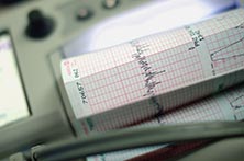
Electrocardiography (EKG or ECG) is a technique used to monitor a person’s cardiovascular activity in real-time. It is a vital part of medical practices, and we believe that it is equally important in dental practices. Our dentists are trained in the interpretation of EKGs, as well as emergency medical care.
We do not believe that it is possible to be too cautious, or too safety conscious. Therefore, we go to great lengths to prepare for any possible emergency or complication – then we go to even greater lengths to ensure that problems do not happen.